Nursing Care Plan

Search Here

NCP Hydrocephalus : Acute Pain and Ineffective Cerebral Tissue Perfusion

Hydrocephalus is a buildup of fluid inside the skull, leading to brain swelling. Hydrocephalus is caused by cerebrospinal fluid flow problems, the fluid that surrounds the brain and spinal cord. This fluid carries nutrients to the brain, eliminating waste from the brain, and acts as a cushion.

CSF normally moves through the area of the brain called ventricles, around the outside of the brain and spinal cord. This fluid is then absorbed into the bloodstream. Fluid buildup can occur in the brain if the flow or absorption is blocked or if too much fluid is produced. Accumulation of fluid puts pressure on the brain, pushing the brain to the skull and damaging or destroying brain tissue.


Hydrocephalus - Nursing Diagnosis and Interventions (NIC - NOC)


1. Ineffective cerebral tissue perfusion related to the increased volume of cerebrospinal fluid.

NOC: Circulation status

Expected outcomes (NOC):

1. Shows the status of circulation which is characterized by the following indicators:
  • Systolic and diastolic blood pressure within the expected range.
  • No orthostatic hypotension.
  • No noisy large blood vessels.

2. Demonstrate the cognitive abilities which is characterized by the following indicators:
  • Communicate clearly and in accordance with the age and ability.
  • Show attention, concentration and orientation.
  • Shows the long-term memory and the present.
  • Process information.
  • Making the decision properly.

NIC Intervention

Monitor  :

1. Vital signs.
2. Headache.
3. The level of awareness and orientation.
4. Diplopia, nystagmus, blurred vision, visual acuity.
5. Monitoring ICT
  • ICT monitoring and neurological response of patients to treatment activities.
  • Monitor the tissue perfusion pressure.
  • Note the change in the patient's response to a stimulus.
6. Management of peripheral sensation
  • Monitor for parestesis: numbness or tingling.
  • Monitor fluid status, including intake and output.

Collaborative Activity

1. Maintain the thermodynamic parameters within the recommended range.
2. Give medicines to increase intravascular volume, according to the request.
3. Give the drugs that cause hypertension to maintain cerebral perfusion pressure, according to the request.
4. Elevate the headboard of 0 to 45 degrees, depending on the patient's condition.


2. Acute Pain related to an increase in ICT

NOC:

1. Pain Level
  • Reports of pain.
  • Frequency of pain.
  • The duration of pain.
  • Facial expressions to pain.
  • Anxiety.
  • Changes in vital signs.
  • Changes in pupil size.
2. Pain Control
  • Mention the factors that cause.
  • Mention the time of the pain.
  • Analgesic use as indicated.
  • Mention the painful symptoms.

NIC:

1. Pain Management
  • Show overall assessment of pain including the location, characteristics, duration, frequency, quality, intensity and pain predisposing factors.
  • Observation of non-verbal cues of discomfort, especially if it can not communicate effectively.
  • Ensure patients receive appropriate analgesic.
  • Determine the impact of pain on quality of life (eg; sleep, activity, etc.).
  • Evaluation with the patient and health care team, the effectiveness of the control of pain in the past used.
  • Assess the patient and family to seek and provide support.
  • Provide information about pain, for example; cause, how long will expire and the anticipation of discomfort from the procedure.
  • Control of environmental factors that may influence a patient's response to discomfort (eg, room temperature, light and noise).
  • Teach for using non-pharmacological techniques (eg relaxation, guided imagery, music therapy, distraction, etc.).

Nursing Diagnosis for Morbid Obesity

Nursing Care Plan for Morbid Obesity

Obesity is defined as having excess fat in the body. Obesity increases the risk of other diseases, such as diabetes and high blood pressure. Doctors use the BMI (body mass index), which is based on weight and height to determine whether you suffer from obesity.

Extreme obesity or severe obesity known as morbid obesity. Morbid obesity is a condition where a person has a BMI over 40 or more.

Symptoms associated with obesity include:
  • Hard to sleep.
  • Snoring.
  • Stop breathing for a while suddenly during sleep.
  • Back pain or joint.
  • Excessive sweating.
  • Always feel hot.
  • Rash or infection of the skin folds.
  • Difficulty breathing.
  • Often sleepy and tired.
  • Depression.

There are genetic and hormonal influences on body weight. The most fundamental thing is that obesity occurs when the body receives more calories rather than burn it. Calories are then accumulate and become fat.

Obesity is usually the result of a combination of the following factors:
  • Not physically active so that the burning of fat becomes a little.
  • Eating high-calorie foods, especially fast food.
  • Some women difficult to lose weight after giving birth, it triggers obesity.
  • Lack of sleep.
  • Certain drugs, such as diabetes drugs, anti-seizure, antidepressants, antipsychotic, steroids and beta blockers.
  • Other medical problems.


Nursing Diagnosis for Morbid Obesity
  1. Imbalanced Nutrition: more than body requirements related to the increase in the intake of nutrients.
  2. Ineffective breathing pattern related to a decrease in lung expansion.
  3. Diarrhea related to changes in dietary fiber.
  4. Impaired tissue perfusion related to immobilization or bed rest.
  5. Disturbed Body Image or Low Self-Esteem related to psychosocial factors (emphasis to lose weight).
  6. Activity intolerance related to being overweight.
  7. Excess fluid volume relate to excess intake of sodium / fluid.

Disturbed Body Image related to Rheumatoid Arthritis

Nursing Care Plan for Rheumatoid Arthritis

Rheumatoid arthritis is a disease which has long been known and spread throughout the world and involving all races and ethnic groups. Rheumatoid arthritis is often found in women, with the ratio of women to men is 3: 1. The tendency for women suffering from rheumatoid arthritis and remissions are common in women who are pregnant, this raises the suspicion of the presence of hormonal balance factor as one of the factors that affect this disease.

Most patients show symptoms of chronic intermittent disease, which if left untreated will cause damage to joints and progressive joint deformity that causes disability and even premature death.

Disturbed Body Image related to changes in the ability to perform the duties of a general, an increase in energy use, the imbalance of mobility.

Evidenced by:
  • Changes in the function of diseased body parts.
  • Focus on past strength and appearance.
  • Changes in lifestyle / physical ability to continue the role, job loss, dependence on the nearest person.
  • Changes in social engagement; a feeling of isolation. Feelings of helplessness, hopelessness.

Expected outcomes: The patient will :
  • Expressing increased confidence in the ability to cope with illness, changes in lifestyle, and possible limitations.
  • Develop a realistic plan for the future.

Intervention and Rationale

1. Encourage disclosure about the problem of the disease process, hope for the future.
R /: Give the opportunity to identify the fear / misconceptions and deal with it directly.

2. Discuss the meaning of loss / change in patients / people nearby. Ascertain how the patient's personal views on the functioning of day-to-day lifestyle.
R /: Identify how the disease affects self-perception and interaction with others will determine the need for intervention / counseling further.

3. Discuss the patient's perception of how the people closest to accept limitations.
R /: Gestures verbal / non-verbal people nearby can have a major influence on how the patient sees himself.

4. Acknowledge and accept the feelings of the bereaved, hostile, dependency.
R /: constant pain would be exhausting, and feelings of anger and hostility are common.

5. Pay attention to the behavior of withdrawing, denying use or too noticed a change.
R /: Can demonstrate emotional or maladaptive coping methods, requiring further intervention.

7. Involve patients in the treatment plan and schedule of activities.
R /: Increase the feeling of self-esteem, encourage independence, and encourage participation in therapy.

8. Assist in need of care required.
R /: Maintaining the appearance that can improve self-image.

9. Provide positive support when necessary.
R /: Allows patients to feel good about themselves. Reinforcing positive behavior. Increase confidence.

10. Collaboration: Refer to psychiatric counseling, eg specialist psychiatric nurse, psychologist.
R /: Patient / person nearby may need support for dealing with long-term process / incapacity.

11. Collaboration: Give medicines as directed, eg; antianxiety drugs and mood enhancer.
R /: It may be required at the time of the advent of the Great Depression, until the patient increases coping abilities more effectively.

Gastroesophageal Reflux Disease (GERD) - Assessment and Physical Examination

Gastroesophageal Reflux Disease (GERD) - Assessment

1. General condition
Covering conditions such as the level of tension / fatigue, qualitative level of consciousness or GCS and client verbal response.

2. Vital signs
Includes examining:
Blood pressure: should be examined in a different position, assess the pulse pressure, and pathological conditions.
Pulse rate.
Respiratory rate.
Temperature.

3. Main complaint
Assessed onset, duration, quality and characteristics, severity. Location, precipitating factors, related manifestations:
Typical complaints (esophagus): heartburn, regurgitation, and dysphagia.
Atypical Complaints (extra esophagus): chronic cough, hoarseness, pneumonia, pulmonary fibrosis, bronchiectasis, and non-cardiac chest pain.
Another complaint: weight loss, anemia, hematemesis or melena, odynophagia.

4. Past medical history/Previous health
Other gastrointestinal diseases.
Drugs that affect gastric acid.
Allergy / immune response reaction.

5. Family medical history


Physical Examination

1. General condition: The general condition may include an impression of illness including facial expressions and patient positioning, awareness may include qualitative assessment.

2. Examination of vital signs: pulse (frequency, rhythm, quality), blood pressure, respiration (frequency, rhythm, depth, respiratory pattern) and body temperature.

3. Examination of the skin, hair and lymph nodes.
Skin: The color (pigmentation, cyanosis, jaundice, pale, erythema and others), turgor, skin moisture and the presence / absence of edema.
Hair: color, luxuriance, distribution and other characteristics.
Lymph nodes: the shape and signs of inflammation that can be assessed in the anterior cervical region, inguinal, occipital and retroauricular.

4. Examination of the head and neck
Head: the shape and size of the head, the hair and scalp, the crown of the head (fontanelle), seen from the eyes of vision, palpebrae, eyebrows eyelashes, conjunctiva, sclera, pupil, lens,
Ear: ear canal, tympanic membrane, mastoid, auditory acuity,
Nose and mouth: lips, gums, tongue, salivation.
Neck: Neck stiffness, size, shape, position, consistency, and presence or absence of pain swallow.

5. Chest examination: Examination of the chest is the organ of the lung and heart. In general, the form asked the chest, lungs state which includes symmetric, the movement of the breath.

6. Examination of the abdomen: about the size or shape of the abdomen, abdominal wall, bowel, abdominal wall tension or tenderness, and palpation in the liver, spleen, kidneys, bladder that determined the presence or absence and the enlargement of the organ, then examination of the anus, rectum and genetal.

7. Examination of the limbs and neurologic: checked for range of motion, balance and gait, the grip, the leg muscles, and others.

Chronic Pain related to Stomach Cancer


Nursing Care Plan for Stomach Cancer

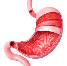
Stomach Cancer is a disease that occurs in the stomach. The average age of patients with stomach cancer is 45 years and above. However there are cases where the disease is under the age of 45 years.Like most other types of cancer, stomach cancer cause is not known with certainty.

But according to some experts, the cause of stomach cancer is suspected because of the bacterium Helicobacter pylori, less healthy eating patterns, consuming less vegetable fiber, or fruit and unhealthy lifestyles, such as smoking, alcohol consumption and eating food that is burned.

In the early stages, the symptoms of gastric cancer is not clear. This can lead to gastric cancer patients often come to the doctor in a state of advanced stage. Symptoms at this stage may include a burning sensation in the pit of the stomach, weight loss and discomfort in the upper abdomen.

While symptoms at an advanced stage may include pain in the upper abdomen, nausea, vomiting, drastic weight loss, vomiting of blood, black bowel, anemia and accompanied by symptoms spread in other organs. For example, an enlarged liver, jaundice, enlarged abdomen due to accumulation of fluid or ascites, and so forth.


Nursing Diagnosis : Chronic Pain related to irritation of the mucosa of the esophagus, surgical response.

Goal: Pain is reduced or adapted.

Expected outcomes:
  • Subjectively say the pain is reduced or adapted.
  • Pain scale 0-2.
  • Vital signs within normal limits.
  • The face looks relaxed.

Interventions and Rationale :

1. Explain and help the patient with pain relief measures, non-pharmacological and noninvasive.
R/ : Approach by using relaxation and nonpharmacological therapy has demonstrated effectiveness in reducing pain.

2. Assess pain with PQRST approach.
R/ : PQRST approach can comprehensively assess the condition of the patient's pain. If the patient experiences pain scale 3 (on a scale of 0-4), this is a warning that needs to be alert because the clinical manifestations of postoperative complications esophagectomy.

3. Rest the patient when pain appears.
R /: Rest, physiologically reduces the need for oxygen required for basal metabolic needs.

4. Encourage deep breathing relaxation techniques when pain appears.
R /: Increase the intake of oxygen, thereby decreasing pain, secondary to intestinal ischemia.

5. Teach technique of distraction during painful.
R /: Distraction (diversion) can reduce internal stimulation.

6. Treating patients in intensive care.
R /: In order to control the patient's pain must be treated in intensive care. Quiet environment will reduce external pain stimulus. Restrictions on visitors to help improve the condition of the room oxygen which would be reduced if a lot of visitors who are in the room. Rest will decrease the oxygen demand of peripheral tissues.

7. Perform touch management.
R /: Touch management - a touch of psychological support - can help reduce pain.

8. Increase knowledge of patients regarding the causes of pain and develop how long the pain will last.
R /: Knowledge will help reduce the pain and can help develop a plan patient adherence to therapy.

Hyperthermia related to Neonatal Sepsis


Nursing Diagnosis and Interventions for Neonatal Sepsis

Sepsis is a syndrome characterized by clinical signs and symptoms of severe infection that can progress toward septicemia and septic shock. (Doenges, 1999)

While neonatal sepsis is a severe infection that affects neonates with systemic symptoms and there are bacteria in the blood. Neonatal sepsis course of the disease can take place quickly so often not monitored, without adequate treatment babies can die within 24 to 48 hours. (Surasmi, 2003).


Nursing Diagnosis and Interventions for Neonatal Sepsis

Hyperthermia related to damage control temperature, secondary to infection or inflammation.

Expected outcomes:
  • The body temperature within normal limits.
  • Pulse and breathing frequency within normal limits.

Intervention and Rationale:

1. Monitoring of vital signs every two hours and monitor skin color.
R /: Changes in vital signs that would significantly affect the regulatory processes or metabolism in the body.

2. Observation of seizures and dehydration.
R /: Hyperthermia potential to cause seizures that will worsen the patient's condition and can cause the patient to lose a lot of fluid in the evaporation of an unknown number and can cause the patient goes into a state of dehydration.

3. Give compress with warm water in the axilla, neck and groin, avoid using alcohol to compress.
R /: Compress the axilla, neck and groin are large blood vessels, which helps reduce fever. The use of alcohol is not done because it will cause a decrease and an increase in heat drastically.

4. Collaboration: Give antipyretics as needed if the heat does not go down.
R /: Giving antipyretics are also required to reduce the heat immediately.


Nursing Care Plan for Hyperthermia

Psychotherapy of Anxiety Disorders

Anxiety, worry, fear, is a common psychological symptoms and can be felt by each individual. Anxiety reactions usually occur frequently in adults, but children can also face the anxiety, such as temporarily abandoned by parents, first day of school or at the time wanted the exam. In the case of the elderly, the reaction of anxiety often occur when they face the pressure (stress) with the difficulties that can be faced and the difficulties that can not be faced, such as work pressure, pressure at school / college, the pressure on the issue of romance as well as pressure on health issues.

Excessive anxiety reaction and settled continuously over a considerable period of time can turn into a disorder, which anxiety disorder. The nature of anxiety disorders can produce a response to the physical and psychological.

Anxiety disorder is a serious mental illness characterized by feelings of great anxiety and excessive, such as feelings of excessive fear, heart pounding harder, shortness of breath, sweating, breathing short, easy to feel dizzy and feeling uneasy.

People who have anxiety disorders often experience these reactions and more severe, causing them distress and causes them unable to perform their daily work. They become very alert, because it is afraid of danger, as a result they are difficult to relax and also difficult to feel at ease in many situations.

Anxiety - Definition According to Experts

Anxiety - Definition According to Experts

According to Freud (in Alwisol, 2005: 28) says that anxiety is the ego function to warn people about the possibility of a danger, so it can be prepared the appropriate adaptive response. Anxiety serves as a mechanism that protects the ego because the anxiety signal to us that there is a danger and if not done the right thing, the danger will increase to ego defeated.

Lefrancois (1980) states that anxiety is an unpleasant emotional reaction, which is characterized by fear. However, according to Lefrancois, the anxiety danger is ambiguous, for example there is a threat, given the barriers to personal desires, their depressed feelings that arise in consciousness.

The concept of anxiety plays a very fundamental theories of stress and adjustment (Lazarus, 1961). According to Post (1978), anxiety is an unpleasant emotional state, which is characterized by subjective feelings like stress, fear, worry, and also characterized by active central nervous system. Freud (in Arndt, 1974) describes and defines anxiety as an unpleasant feeling, followed by certain physiological reactions such as changes in heart rate and breathing. According to Freud, anxiety involves the perception of unpleasant feelings and physiological reactions, in other words, anxiety is a reaction to a situation which is considered dangerous.

Not much different from Lefrancois opinion is the opinion expressed by Johnston (1971) which states that anxiety can occur because of disappointment, dissatisfaction, insecurity or the hostility with others. Kartono (1981) also revealed that neurosa anxiety is psychological condition in chronic fear and anxiety, though there is no specific stimuli. According Wignyosoebroto (1981), there is a fundamental difference between anxiety and fear. On fear, what is the source of the cause can always be designated significantly, whereas the source of anxiety cause can not be appointed to the firm, clear and precise.

Maramis (1995) states that anxiety is a tension, insecurity, fear, which arises because perceived will experience unpleasant incident.

Lazarus (1991) states that anxiety is the individual's responses to what they're getting. Anxiety is a feeling that is painful, such as anxiety, confusion, etc., associated with the subjective aspect of emotion. Anxiety is a common symptom at this point, because it is along the journey of human life, from birth until the death, anxiety is often present.

Saranson and Spielberger (in Darmawanti 1998) states that anxiety is a reaction to an experience for individuals perceived as a threat. Anxiety is a feeling of uncertainty, panic, fear, without knowing what is feared and can not eliminate the feelings of anxiety and the anxiety.

Hyperthermia and Acute Pain - NCP for Mastoiditis

Nursing Care Plan for Mastoiditis

Hyperthermia and Acute Pain - NCP for Mastoiditis
Mastoiditis is an inflammation of the mastoid bone, usually from the tympanic cavity. The expansion of middle ear infections repeatedly can cause changes in the mastoid, such as thickening of the mucosa and accumulation of exudate. Over time there is inflammation of the bone (osteitis) and collecting exudate / pus that more and more, eventually finding a way out. The weak areas are usually located behind the ear, causing an abscess superiosteum.

According to George (1997: 106), the clinical manifestations in patients with mastoiditis include:
  • The fever usually disappear and arise.
  • Pain tends to settle and throbbing, located around and inside the ears, and experience tenderness in the mastoid.
  • Hearing loss.
  • Tympanic membrane bulging contain skin that has been damaged and discuss sebaceous (fat).
  • Posterior canal wall hanging.
  • Postauricular swelling.
  • A large discharge through the ear canal and the odor.


Nursing Diagnosis and Interventions for Mastoiditis

1. Acute Pain is related to inflammation of the mastoid bone because of infection.

Goal: Pain is resolved.

Expected outcomes:
  • Pain is reduced.
  • Pain scale decreased.
  • The face looked relaxed.
Interventions :

1. Review the scale of pain, location, intensity.
R /: Knowing the effectiveness of interventions.

2. Provide a comfortable position.
R /: Reduce pain.

3. Teach relaxation techniques and create a tranquil environment.
R /: Turning his attention to the pain and reduces pain.

4. Collaboration of analgesics, antibiotics, and anti-inflammatory as indicated.
R /: It can reduce pain, kill germs and reduce inflammation and accelerating healing.


2. Hyperthermia related to the inflammatory process.

Goal: The body temperature may be normal (36 0- 37 0 C)

Expected outcomes:
  • The body temperature within normal range (36 0-37 0 C).
  • The skin does not feel warm.
  • The face does not look red.
  • Prevent dehydration.
Interventions :

1. Monitor the input and output.
R /: To find out the patient's fluid balance.

2. Measure the temperature every 4-8 hours.
R /: To determine the condition of the client's body temperature.

3. Teach warm compresses, and a lot of drinking
R /: To reduce body heat and replace lost body fluids.

4. Collaboration with the administration of antipyretics.
R /: To reduce the heat.

Possible Nursing Diagnosis for Trigeminal Neuralgia

Possible Nursing Diagnosis for Trigeminal Neuralgia
Trigeminal Neuralgia

Neuralgia is a stabbing pain that arises occasionally, but short and heavy, which occurs along the distribution of a nerve. Trigeminal neuralgia (NT) is neuralgia on the trigeminal nerve (fifth cranial nerve) that is responsible for sensation in the face. Trigeminal neuralgia (facial pain) is characterized by brief episodes of strong facial pain, stabbing, and like electricity.

According to Dr. Dito Anurogo, Trigeminal Neuralgia is a complaint of pain attacks one side of the face are repeated. Called trigeminal neuralgia, because facial pain occurs in one or more nerves than the three branches of Trigeminal nerve. This large nerve located in the brain and carries sensation from the face to the brain. The pain is caused by a disturbance in Trigeminal nerve function in accordance with the regional distribution of innervation of one branch of the trigeminal nerve caused by a variety of causes.

Etilogy trigeminal neuralgia is still not fully understood. There is one theory that because the blood vessels, especially the superior cerebral artery, into decompression, so that chronic irritation of the trigeminal nerve into the root section. This irritation causes increased afferent controls blame, or sensory nerves. Risk factors that can trigger is multiple sclerosis and hypertension. Other factors that may cause neuralgia including herpes virus infections, infections of the teeth and jaws, and brain stem infarction. (Miller, 2009 in Lewis 2011).

Nursing Diagnosis that may appear on the client with Trigeminal Neuralgia according Muttaqin, Arif (2010) and Ackley, Betty J., Gail B. Ladwig (2013) is as follows.
  1. Pain (acute / chronic) r / t trigeminal nerve compression and inflammation of the temporal artery.
  2. Imbalanced Nutrition  : Less than Body Requirements r / t pain during chewing.
  3. Ineffective individual coping r / t severe pain, excessive threat to the self-alone.
  4. Knowledge Deficit: on the condition and needs medication r / t cognitive limitations.
  5. Anxiety r / t prognosis of disease and changes in health.
  6. Ineffective management of therapeutic regimen r / t less knowledge about the prevention of stimulus triggers pain.
  7. Risk for injury to the eyes r / t the risk factors: possible reduction in corneal sensation.

Disturbed Body Image NCP for Dermatitis

Nursing Care Plan for Dermatitis

Disturbed Body Image NCP for Dermatitis
Dermatitis is inflammation of the skin. Dermatitis can have many causes and occurs in many forms. Dermatitis usually involves an itchy rash on swollen, reddened skin.

Dermatitis is a common condition that's not contagious and usually isn't life-threatening. Even so, it can make you feel uncomfortable and self-conscious.

Disturbed Body Image related to the appearance of the skin that is not good.

Goal: Development of an increase in self-acceptance.

Expected outcomes:
  • Develop an increase in the willingness to accept a state of self.
  • Follow and participate in self-care measures.
  • Reported feeling in control of the situation.
  • Reinforces the positive support of the self-governing.
  • Express attention to self-healthier.
  • Seemed not to notice the condition.
  • Using a technique to hide flaws and emphasize techniques for improving the appearance.

Interventions :

1. Assess the patient's self-image disturbance in (avoiding eye contact, self-deprecating speech, expression sick state of the condition of the skin).
R /: Disturbed self-image will accompany any disease or condition were apparent to the patient. Impression of someone against itself will affect the self-concept.

2. Identify the psychosocial stages of development stages.
R /: On the relationship between the stages of development, as well as the self-image and understanding of the patient's reaction to the skin condition.

3. Provide an opportunity for disclosure. Listen (by way of an open, non-judgmental) to express mourning / anxiety about body image changes.
R /: Patients in need of the experience that must be listened to and understood.

4. The sense of concern and fear of patients. Help patients who are anxious to develop the ability to assess themselves and identify and resolve problems.
R /: This action provides an opportunity for health workers to neutralize unnecessary anxiety and restore the reality of the situation. Fear is a destructive element patient adaptations.

5. Encourage socialization with other people.
R /: Increase self-acceptance and socialization.

Definition of Hypertension According to the Experts

Definition of Hypertension According to the Experts
Hypertension is one of the cardiovascular system diseases that are often found in the community. Hypertension is not a contagious disease, but it should always be wary. High blood pressure or hypertension and arteriosclerosis are two basic conditions that underlie many forms of cardiovascular disease.

Furthermore, high blood pressure also causes kidney disorders. Until now, efforts to both prevent and treat hypertension has not been entirely successful, because of the inhibiting factors such as lack of knowledge about hypertension (understanding, signs and symptoms, causation, complications) and also treatment.

Various factors play a role in this case one of them is a modern lifestyle. Selection of fatty foods, unhealthy activity habits, smoking, drinking coffee are some of the things that is suspected as a factor that contributes to this hypertension. This disease can be the result of modern lifestyles and can also be a cause of various non-infectious diseases.

To know more about this disease and to know, then we will discuss about hypertension.


Definition of Hypertension According to the Experts


Hypertension was defined as systolic blood increase greater than or equal to 140 mmHg or diastolic blood pressure greater than or equal to 90 mmHg (Anindya, 2009).


Hypertension is defined as blood pressure persistent, where the systolic pressure above 140 mmHg and diastolic pressure above 90 mmHg. (Tom Smith, 1995)

Hypertension is the increase in systolic blood pressure over 140 mmHg and diastolic blood pressure over 90 mmHg (Luckman Sorensen, 1996).

Hypertension was defined by the Joint National Committee on Detection (JIVC) as pressure higher than 140/90 mmHg and classified according to the degree of severity, ranging from blood pressure (BP) high normal to malignant hypertension.

High blood pressure or hypertension is a condition a person's blood pressure is at levels above normal. And the consequences of this situation is the emergence of diseases that interfere with the patient's body. In hypertensive disease is a health problem and need of prevention. (Sudjaswandi: 2002 h 17)

Hypertension is defined as blood pressure that persistent systolic pressure above 140 mmHg and diastolic above 90 mmHg. The aging population, hypertension is defined as systolic pressure of 160 mmHg and a diastolic pressure of 90 mmHg. (Smeltzer, 2001).

Hypertension is defined as systolic blood pressure of 140 mmHg or a diastolic pressure of at least 90 mmHg. Traditional terms of hypertension "mild" and "moderate" failed to explain the influence of the major high blood pressure in cardiovascular disease. (Anderson: 2006 h 582)

Hypertension is high blood pressure or medical terms, explain hypertension is a condition where an interruption in blood pressure regulation mechanism (Mansjoer, 2000: 144)

Categorized as mild hypertension if diastolic pressure between 95-104 mmHg, moderate hypertension if diastolic pressure between 105 and 114 mmHg, and severe hypertension if diastolic pressure 115 mmHg or more. This division is based on the increase in the diastolic pressure is considered more serious because of the increase in systolic (Tom Smith, 1995).

Hypertension is systolic pressure exceeds the settled state of 140 mmHg or diastolic pressure higher than 90 mmHg. This diagnostic can be ascertained by measuring the average blood pressure at two separate times (School of Medicine, 2001: 453)

Nursing Care Plan for Trachoma

Nursing Care Plan for Trachoma
Trachoma is the world's leading cause of preventable blindness and the second cause of blindness after cataract. Blindness from trachoma occurs after years of repeated infection with the microorganism, Chlamydia trachomatis. The process of infection and re-infection starts in early childhood and may continue to adulthood, if the cycle is not broken.

Women have a two to three times the rate of advanced trachoma and blindness than men, because as mothers, grandmothers and older sisters who care for children (the main source of active trachoma infection), they are redundant and are constantly exposed to bacteria.

Trachoma is caused by Chlamydia trachomatis and is spread through direct contact with the eyes, nose, and throat are exposed to liquid (containing bacteria) of people living with, or in contact with inanimate objects, such as towels and / or rags, which once contact is similar to the liquid , Flies can also be a route of transmission. If left untreated, repeated trachoma infection can result in entropion which is a form of permanent blindness and accompanied by pain if the eyelid turns inward, as this causes the eyelashes to scratch the cornea. Children are most susceptible to this infection because of their tendency to easily get dirty, but the effects of blurred vision and other more severe symptoms are often not felt until adulthood.

These bacteria have an incubation period of 5 to 12 days after a person experiences symptoms of conjunctivitis, or irritation similar to "pink eye." Endemic trachoma blindness is the result of several episodes of reinfection that produces continuous inflammation of the conjunctiva. Without reinfection, the inflammation will be gradually improved.

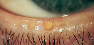
Inflammation of the conjunctiva is called "active trachoma" and usually seen in children, especially children of pre-school (elementary). It is characterized by white bumps on the bottom surface of the upper eye lid (conjunctival follicles or germinal centers of lymphoid). Non-specific inflammation and thickening often associated with papillae. Follicles may also appear at the junction of the cornea and sclera (limbal follicles). Active trachoma will often be irritating and have a watery fluid. Secondary bacterial infection may occur and cause a purulent discharge.

Further symptoms include:
Dirty discharge from the eyes - not tears (emissions or secretions containing mucus and pus from the eyes).
Swelling of the eyelids.
Trichiasis (turned eyelashes).
Swollen lymph nodes in the front of the ear.
The appearance of lines scarring of the cornea.
Complications in the ear, nose and throat.

The main complication is the most important or ulcers (sores / irritations) on the cornea due to a bacterial infection.


Nursing Diagnosis for Trachoma
  1. Acute pain: eye related to swelling of the lymph nodes, photophobia and inflammation.
  2. Disturbed Sensory Perception: Visual related to damage to the cornea.
  3. Risk for infection, the spread related to lack of knowledge.
  4. Body image disorders related to loss of vision.

Risk for Impaired Skin Integrity - NCP for Dysentery

Nursing Care Plan for Dysentery

Dysentery is derived from the Greek, ie dys (= disorder) and enteron (= intestine). so dysentery
is a gastrointestinal disease in the form of intestinal infection or inflammation of the intestines caused by bacteria, which causes severe diarrhea. Each individual course of the disease is more varied, with some people suffering from dysentery have mild symptoms, while others may experience severe diarrhea with or without vomiting which can pose a risk of dehydration. Fortunately dysentery can be easily treated with antibiotics and antiparasitic drugs. Dysentery if untreated can lead to severe dehydration.

The most common cause of dysentery and is often found in people are not washing hands after using public restrooms or not washing hands before eating. Indeed simple enough to cause dysentery as a classic case, but that the reality is often the case. Broadly speaking, the cause of dysentery is closely related to the cleanliness of our surroundings and clean living habits.

Symptoms that arise in dysentery, among others:
  • Time dysentery symptoms can last between 5-7 days or even longer.
  • Patients experiencing abdominal cramps (colic).
  • Patients experiencing pain during bowel movements (tenesmus).
  • Defecation accompanied by mucus.
  • Defecation with bloody stools.
  • High fever (39.5 to 40 degrees Celsius).
  • Vomiting.
  • Anorexia.
  • Sometimes accompanied by symptoms like encephalitis and sepsis
  • (seizures, headache, lethargy, stiff neck, hallucinations).

Risk for Impaired Skin Integrity : perianal related to an increase in the frequency of bowel movements (diarrhea).

Goal: skin integrity is not compromised.

Expected outcomes:
  • No irritation: redness, blisters, hygiene maintained.
  • Families are able to demonstrate perianal care of properly.
Intervention:
1) Discuss and explain the importance of keeping the bed.
R / Hygiene prevent the proliferation of germs.

2) Demonstrate and involve families in caring for perianal (when wet clothing and replace the bottom as well as the base).
R /: Preventing skin iritassi unexpected because humidify and stool acidity.

3) Position the bed or sitting at intervals of 2-3 hours
R /: Smooth vascularity, reducing the pressure for a long time, so there is no ischemia, and irritation.

Imbalanced Nutrition : Less than Body Requirements - NCP Diabetic Ketoacidosis


Nursing Care Plan for Diabetic Ketoacidosis

Diabetic Ketoacidosis (DKA) is a potentially life threatening condition of diabetes occurring mostly in patients with Type 1 diabetes but on occasion also in patients with Type 2 diabetes. This disease is a potentially fatal complication of diabetes that occurs when insulin levels are far lower than what the body needs.

The most frequent early signs of Diabetic Ketoacidosis or DKA is a sharp increase in polydipsia and polyuria. Whilst Polydipsia means excessive thirst, Polyuria refers to the excessive production of dilute urine. Other symptoms include malaise, generalized weakness, and tiredness. These may then progress if untreated to vomiting, deep gasping breathing (kussmaul respiration), dehydration, rapid weight loss in patients newly diagnosed with Type 1 diabetes, mild disorientation, confusion and occasionally coma.


Imbalanced Nutrition : Less than Body Requirements related to the insufficiency of insulin, decreased oral input, hyper-metabolic status.

Expected outcomes: The client will;
  • Digesting the number of calories / nutrients right.
  • Shows the energy level normally.
  • Demonstrating steady weight or adding appropriate normal range.

Interventions :

1. Monitor weight daily or as indicated.
R:/ Assessing adequate food intake, including absorption and utilities.

2. Determine the diet and eating patterns of patients and compare it with the food being spent.
R:/ Identify deficiencies and deviations from the therapeutic needs

3. Auscultation bowel sounds, note the presence of abdominal pain / abdominal bloating, nausea, vomit undigested food, keep fasting as indicated.
R:/ Hyperglycemia and disorders of fluid and electrolyte balance may decrease motility / function of the stomach (distention or paralytic ileus) that will affect the choice of intervention.

4. Give foods that contain nutrients then try giving a more solid that can be tolerated.
R:/ Oral feeding is better if the patient is conscious and good gastrointestinal function.

5. Involve patients in planning family as indicated.
R:/ Provide information on the family to understand the nutritional needs of the patient.

6. Observation of signs of hypoglycemia.
R:/ Hypoglycemia can occur because of a reduced carbohydrate metabolism while still given insulin, it can potentially be life threatening and should be recognized.

Collaboration:
1. Monitor proofing acetone, pH and HCO3.
R:/ Monitor the effectiveness of insulin in order to stay in control.

2. Give regular insulin treatment as indicated.
R:/ Facilitate the transition on carbohydrate metabolism and lowers the incidence of hypoglycemia.

3. Examination of blood sugar.
R:/ Monitor blood sugar is more accurate than the reduction of urine to detect fluctuations.


NCP for Congenital Heart Disease : Assessment, Nursing Diagnosis and Interventions


Congenital Heart Disease Nursing Diagnosis and Interventions
Nursing Care Plan for Congenital Heart Disease

Congenital Heart Disease (CHD) is a heart disease which is inborn, because it occurs when a baby still in the womb. At the end of the seventh week of pregnancy, heart formation is complete; so the formation of cardiac abnormalities occur in early pregnancy. Causes of Congenital Heart Disease (CHD) often can not be explained, although several factors are considered as a potential cause (Rahayoe, 2006).

Congenital heart defects is heart defects or malformations that appear at birth, in addition to congenital heart disease is a disorder of the heart anatomy brought from conception to birth. Most congenital heart defects include structural malformations in the heart and major blood vessels, both the left and that leads to the heart (Nelson, 2000). This disorder is the most common congenital abnormalities in children, about 8-10 of 1,000 live births.

This congenital heart defect does not always give symptoms shortly after birth, it is not uncommon these disorders has been discovered after a few months old, or even found after a few years old. This disorder can be mild so as not detected at birth. However, in particular children, the effects of this disorder is so severe that a diagnosis was enforceable even before birth. With the sophistication of medical technology in the field of diagnosis and treatment, many children with congenital heart defects can be remedied and well into adulthood (Ngustiyah, 2005).

The cause of congenital heart disease can not be known with certainty, but there are several factors that have an influence on the expected increase in the incidence of CHD.

These factors are:
1. Prenatal factors:
  • Mothers suffering from infectious diseases: rubella.
  • Maternal alcoholism.
  • Maternal age over 40 years.
  • Peyakit mother suffering from diabetes mellitus who require insulin.
  • Mothers taking sedative drugs or herbs.
2. Genetic factors
  • Children born before suffering from CHD.
  • Father / mother suffering from congenital diseases.
  • Down syndrome is a chromosomal abnormality example.
  • Born with congenital abnormalities others.

Nursing Care Plan for Congenital Heart Disease

Assessment
  1. Physical assessment (color, pulse, respiration, blood pressure, chest auscultation).
  2. Family history.
  3. Pregnancy history.
  4. Assessment manifestations of congenital heart disease.
  5. Collagen tissue abnormalities.
  6. Complications or consequences of hypoxemia.
  7. Construction of a weak body.
  8. Dyspnea on activity.
  9. Fatigue.

Nursing Diagnosis for Congenital Heart Disease
  1. Risk for decreased cardiac output r / t defect structure.
  2. Altered Growth and Development r / t inadequate oxygen and nutrients to the tissues.
  3. Risk for infection r / t weak physical status.
  4. Altered family processes r / t have children with heart disease.
  5. Risk for injury (complications) r / t cardiac conditions and therapies.


Intervention
  1. Check the blood, red blood cell indices.
  2. Assess the arterial blood gas analysis.
  3. Test oxygen.
  4. Give afterload lowering medications as instructed.
  5. Give diuretic as instructed.
  6. Provide frequent rest periods and sleep periods without interruption.
  7. Encourage quiet activities.
  8. Give a diet high in nutrients, which is balanced to achieve adequate growth.
  9. Monitor height and weight.
  10. Encourage the family to participate in the care process.
  11. Teach families to recognize the signs of complications.

Expected Results
  1. Heart rate, BP and peripheral perfusion are the age-appropriate upper limit of normal.
  2. Exit adequate urine (between 0.5 and 2ml / kg, depending on age).
  3. Children achieve adequate growth.
  4. Families can confront the child with positive symptoms.
  5. Families recognize the signs of complications and take appropriate action.

Disturbed Sensory Perception (visual) related to Blepharitis


Disturbed Sensory Perception (visual) related to Blepharitis
Nursing Care Plan for Blepharitis

Blepharitis or better known as inflammation of the eyelids is one of the eye diseases to watch out for. The disease is caused by the oil glands at the base of the lashes were damaged, causing itching in the eyelids, irritation, even to inflammation. If this is allowed then it would be blepharitis.

Although blepharitis does not cause permanent damage to the eye, but very disturbing activities of our vision.

Symptoms of Blefarits as follows:
  • The eyelids are often greasy.
  • Itch on the eyelids.
  • Burned in the eye.
  • Eyes look red.
  • Frequent watery eyes.
  • Eye swelling of the eyelids.
  • Eyelash gross waking.
  • Very sensitive to light.
  • Have peeling skin around the eyes.
  • Eyelashes fall out.
  • Eyelashes abnormal and irregular direction.

Main causes of the occurrence of blepharitis (inflammation of the eyelids)
  • Infected by the bacteria.
  • Glands damaged eye.
  • Dandruff of the scalp to eyebrows.
  • Rosacea.
  • Allergic to something eg eye drops, eye lens.


Nursing Diagnosis and Interventions for Blepharitis :

Disturbed Sensory Perception (visual) related to reception interference status sensory organs.

Goal: Increase the visual acuity within the limits of individual situations.

Expected outcomes:
  • Know the sensory disturbances and compensate for changes.
  • Identify / fix potential hazards in the environment.

Intervention:

1) Determine the visual acuity, note whether one or both eyes are involved.
Rational: individual needs and choice of interventions varied causes vision loss occurs slowly and progressively.

2) Observe the signs and symptoms of disorientation.
Rational: woke up in unfamiliar surroundings and have limited vision.

3) Orient the patient on the environment, other people in the area.
Rationale: provides increased comfort and familiarity.

4) Notice about blurred vision and eye irritation, which can occur when using eye drops.
Rational: impaired vision / irritation can end 1-2 hours after use of eye drops, gradually decreases with usage.

5) Place the items needed / call bell within reach positions on the near side.
Rational: allowing patients to see objects more easily and facilitate the call for help when needed.

Ineffective Airway Clearance - NCP for Bronchiectasis

Nursing Care Plan for Bronchiectasis

Bronchiectasis is a chronic dilatation of the bronchi and bronchioles that may be caused by various conditions, including lung infections and bronchial obstruction; foreign body aspiration, vomit, and objects from the upper respiratory tract; and the pressure due to a tumor, blood vessels dilated and enlarged lymph nodes (Brunner & Suddart, 2002).

According Suyono (2001) etiology of bronchiectasis are:

1. Infection
Bronchiectasis often occur after a child suffering from pneumonia who frequently relapse and long lasting. Pneumonia is generally a pertussis or influenza complications suffered during the child, pulmonary tuberculosis, and so on.

2. Abnormalities hereditary or congenital abnormalities
In this case bronchiectasis occurs in the womb. Genetic factors or growth factors and fetal development plays an important role. Usually has the characteristics on almost all branches of the bronchi in the lungs one or two. Usually accompanied by other congenital diseases.

3. Bronchial obstruction
Obstruction is meant as a corpus alienum, bronchial carcinoma and other external pressure against the bronchi.


Pathophysiology

According to Brunner & Suddarth (2002) pathophysiology of bronchiectasis starting from infections that damage the bronchial wall, causing loss of supporting structure and produce thick sputum that can eventually clog the bronchi. Bronchial walls become stretched permanently as a result of severe coughing, infection extends to peri bronchial, so that in the case of secular bronchiectasis, each tube is actually dilated pulmonary abscess, which exudates to flow freely through the bronchi. Bronchiectasis is usually local, attacking lung lobe segment. Lower lobe most often affected.

Retention secretion and obstruction resulting eventually cause obstruction distal side alveoli collapse (atelectasis). Due to scarring or fibrosis replaces lung tissue inflammatory reaction that function. At the time the patient suffered respiratory insufficiency with a decrease in vital capacity, decreased ventilation, and an increase in the ratio of residual volume to total lung capacity. There is damage to the gas mixture in the inspiration (ventilation-perfusion mismatch) and hypoxemia.


According Suyono (2001) the signs and symptoms of bronchiectasis as follows:

1. Cough
Hemoptysis is characterized, among others; ongoing chronic productive cough, sputum amount varies, generally polynomial in the morning after there is a sleeping position or wake up from sleep. Sputum consists of three layers:
  • The top layer; rather cloudy, consisting of mucus.
  • The middle layer; clearly consists of saliva.
  • The bottom layer; turbid, consisting of pus and tissue necrosis of bronchial damaged.
2. Hemoptysis
Caused by necrosis or destruction of the bronchial mucosa blood vessels (rupture) and the resulting bleeding.

3. Shortness of breath (dyspnea)
Onset of shortness of breath depends on the extent of bronchiectasis, sometimes causing wheezing sound due to bronchial obstruction.

4. Recurrent fever
Bronchiectasis is a chronic disease, often experience recurrent infection of the bronchi and the lungs, often resulting in fever (recurrent fever).

5. Physical Abnormalities
  • Cyanosis
  • Clubbing
  • Bronchi wet
  • Whezing

Nursing Care Plan for Bronchiectasis

Nursing Diagnosis : Ineffective airway clearance related to the increased production of secretions, thick secretions.

Goal: Maintain a patent airway with breath sounds clean / clear.

Expected outcomes: Demonstrate behaviors to improve airway clearance (effective cough, and issued a secret.)

Intervention:
1. Auscultation of breath sounds and record their breath sounds.
R /: The degree of bronchospasm occurs with airway obstruction and can / not characterized by the presence of breath sounds.

2. Assess / monitor respiratory frequency. Note the ratio of inspiration and expiration.
R /: Tacipneu common to some degree can be found at the reception or during stress / acute infection process. Slowed breathing and elongated compared inspiration expiration frequency.

3. Assess the patient to a comfortable position, Height head of the bed and sat on the back of the bed.
R /: Elevation headboard ease respiratory function by means of gravity. And make it easier to breathe, and help decrease muscle weakness and can be as a tool chest expansion.

4. Help abdominal breathing exercises or lips.
R / To cope with and control of dyspnea and lower air entrapment.

5. Observe the characteristic cough and aid effectiveness action to cough effort.
R /: Knowing the effectiveness of cough.

6. Increase fluid intake till 3000ml / day as tolerated heart and give a warm and fluid intake between as a meal replacement.
R /: Hydration helps to lower the viscosity of secretions, simplify expenditure warm fluids can reduce bronchospasm. Liquids between meals can increase gastric distension and pressures diaphragm.

7. Give the drug as indicated.
R /: Speed up the healing process.

10 Types of Pruritus (Itch)

Pruritus is irritating skin sensation and is characterized by itching, as well as provoking to scratch.

1. Pruritus Gravidarum
Induced by the estrogen hormone, especially in the third trimester of pregnancy, starting from the abdomen or body, then generalized, may be accompanied by symptoms of anorexia, nausea or vomiting are also accompanied by pruritus cholestatic jaundice after 2- 4 weeks due to bile salts in the skin.

2. Cholestatic pruritus
As an expression of cholestatic pruritus sign of obstruction in the bile (obstruction biliarry disease) located in the hepatic region, can also be caused side effects of drugs that provide intra hepatal obstruction resulting billiard acid salt excretion.

3. Senile pruritus
Senile skin dry, easy to suffer from fissures (chapped skin) absolute become pruritic, occurs with or without inflammatory reaction. Itching occurs because light stimulation / temperature changes. The most common area is the external genital area, perineal and perianal.

4. Pruritus on Endocrine System (Diabetes Mellitus, hypoparathyroidism, myxedema)
In DM occurs hyperglycemia, causing irritability of nerve endings in the skin and metabolic glands anogenital region or submammae especially in women.
In hyperparatiroidism an increase in plasma parathyroid hormone resulting in a deficit of calcium in the skin, especially calcium phosphate.

5. Generalized Pruritus
Generalized pruritus occur, especially in the CRF (chronic renal failure) occurs with edema and dry skin (xerosis) due to atrophy of sebaceous glands, and glands sudorifera.
In kidney disease also cause metabolic disorders in phosphorus and calcium, magnesium in serum increased resulting in uremia that cause pruritus, caused by materials that undergo retention, failing kidneys secrete, so need hemodialysis.

6. Pruritus in neoplastic
Pruritus in internal malignancy mainly derived from the lymphoreticular system caused by the incidence of Hodgkin's disease for months, before the symptoms of the underlying disease is known.

7. Pruritus in mycosis fungoides
Is a progressive malignant lymphoma. Pruritic skin lesions arise at the time was not typical and yet there is a malignant infiltration. Pruritus can be settled and intolerant.

8. Pruritus in neurologic
Central nervous deficit / peripheral as a regulator of tactile sensation can cause pruritus.

9. Pruritus in Psychology
Scratching responses differ with pruritus due to other causes. In itch because organic disease there is a correlation between the severity of the itch sensation scratching response. At turns psychological itch scratching response is smaller than the degree of subjective itching, scratching effects seem less and less effect more scratching and picking, and not common sleep disorders.

10. Pruritus in other diseases
  • Gout / arthritis.
  • Hypertension, atherosclerotic cause itching all over the body before the onset apoplexia.
  • Polycythemia veins accompanied by pruritus and urticaria.
  • Fe deficiency, not anemia, due to impaired formation of Fe.

Fatigue Definition

Fatigue

Fatigue is a condition with signs of reduced capacity of a person, for work and reduced efficiency of accomplishment, and this is usually accompanied by a feeling tired and weak. Fatigue can be acute and come on suddenly or chronic and persist. According to other sources of fatigue is a condition of the human body naturally feel tired, which usually happens after physical exercise or mental harm. Fatigue can be acute and come on suddenly or chronic and persist.

Usually after a long exercise, surely people will feel tired, because of all the moving limbs, limbs will be ill and do not want to continue the exercise. However, this fatigue will soon be replaced with good health and well-being. Just like a mix between fatigue and the sense of satisfaction that people feel after working hard in the office or study, this is a healthy and natural fatigue.

Fatigue at the beginning and at the end of pregnancy is also a natural thing, the reason is the increase in the activity of the hormone to be one of the causes as well as fatigue when weight babies in the womb which also makes people pregnant. Tired also can arise from psychological reasons and can be a symptom of certain diseases. But rarely the only symptom of the disease. In undetected diabetes, high levels of sugar than usual and this condition can lead to fatigue. Uncontrolled diabetes can also lead to increased levels of blood sugar and fatigue. In severe anemia, generally dilute blood, heart and lungs should strive to supply oxygen and deliver it to all the rest of the body. Fast heart rate in severe anemia may be accompanied by fatigue, anxiety, fainting, pale biscuits, and shortness of breath.

Nursing Diagnosis and Interventions for Dehydration


Nursing Diagnosis for Dehydration
  1. Fluid volume deficit related to excessive output, less intake.
  2. Risk for ineffective tissue perfusion related to decreased blood flow.
  3. Risk for impaired skin integrity related to decreased skin turgor.
  4. Activity intolerance related to physical weakness.
  5. Risk for Decreased cardiac output related to a decrease in systemic vascular resistance.


Nursing Care Plan for Dehydration

Nursing Interventions for Dehydration

1. Fluid volume deficit related to excessive output, less intake.

Goal: adequate fluid volume so that fluid volume deficiency can be overcome.

Expected outcomes:
  • Maintain fluid balance.
  • Vital signs (pulse = 80-100 beats / min, temperature = 36-37oC)
  • Capillary refill less than 3 seconds.
  • Akral warm.
  • Urine output: 1-2 cc / kg body weight / hour.

Intervention:
  • Monitor vital signs, capillary refill, the status of the mucous membranes.
  • Discuss strategies to stop vomiting and use of laxatives / diuretics.
  • Identification of a plan to increase the optimal fluid balance.
  • Assess the results of the test function electrolyte / kidney.
  • Give / supervise administration of IV fluids.
  • Additional potassium, oral or N as indicated.

2. Risk for ineffective tissue perfusion related to decreased blood flow.

Goal: Maintain / improve tissue perfusion.

Expected outcomes:
  • Vital signs are stable BP = 120/80 mmHg, pulse = 80-100 beats / min, no pale skin.
  • Warm skin.
  • Palpable peripheral pulses.
  • Adequate urine output from 0.5 to 1.5 cc / kg / body weight.
  • CRT is less than 2 seconds.
  • Composmentis consciousness.
  • No chest pain.

Intervention:
  • Assess changes in the level of consciousness, dizziness complaints.
  • Auscultation apical pulse, watch heart rate / rhythm.
  • Assess the skin against the cold, pale, sweating.
  • Record output and urine specific gravity.
  • Observation pale skin, redness, change positions frequently.
  • Keep an eye on pulse oximetry.
  • Give IV fluids as indicated.


3. Risk for impaired skin integrity related to decreased skin turgor.

Goal: Identify and maintain the skin smooth, supple, intact.

Expected outcomes:
  • Good skin turgor, skin intact, no blisters, no redness.

Intervention:
  • Observation reddish, pale.
  • Use skin cream.
  • Discuss the importance of changes in position, it is necessary to maintain the activity.
  • Emphasize the importance of nutrient input / adequate fluid.

Nursing Care Plan for Dehydration

Dehydration

Definition

Dehydration is a condition in which a person who is not fasting experiencing or at risk of dehydration vascular, interstitial or intra-vascular (Sell Lynda Carpenito, 2000: 139).


Nursing Diagnosis for Dehydration
Classification

Classification of dehydration by Donna D. Ignatavicus there are 3 types:

a. Isotonic dehydration
Isotonic dehydration is lost water followed by the electrolyte so that the density remained normal, then this type of dehydration is usually not result in ECF fluid move to the ICF.

b. Hypotonic dehydration
Hypotonic dehydration is the loss of solvent from the ECF exceeds fluid loss, resulting in blood vessels become more concentrated. ECF osmotic pressure decreases, resulting in fluid moves from the ECF to ICF. Vascular volume also decreased, as well as cell swelling occurs.

c. Hypertonic dehydration
Hypertonic dehydration is ECF fluid loss exceeds the solvent is non-osmotic dehydration ECF decreased, resulting in fluid moves from ICF to ECF.


Etiology

Various causes dehydration determine the types of dehydration (According to Donna D. Ignatavicus, 1991: 253).

1. Dehydration
  • Bleeding.
  • Vomiting.
  • Diarrhea.
  • Hypersalivation.
  • Fistula.
  • Ileustomy (cutting intestine).
  • Diaporesis (excessive sweating).
  • Burns.
  • Fasting.
  • Hypotonic therapy.
  • Suction gastrointestinal (stomach wash).
2. Hypotonic Dehydration
  • DM disease.
  • Excess fluid rehydration.
  • Severe and chronic malnutrition.
3. Hypertonic Dehydration
  • Hyperventilation.
  • Diarrhea water.
  • Diabetes Insipedus (ADH hormone decreases).
  • Excessive fluid rehydration.
  • Dysphagia.
  • Impaired thirst.
  • Disorders of consciousness.
  • Systemic infection: increased body temperature.

Clinical Manifestations

Here are the symptoms or signs of dehydration based on its level (Nelson, 2000):
1. Mild dehydration (loss of fluid 2-5% of its original weight)
  • Thirsty, restless.
  • Pulse rate 90 -110 x / minute, normal breath.
  • Normal skin turgor.
  • Urine output (1300 ml / day).
  • Good awareness.
  • Heart rate increased.
2. Moderate Dehydration (loss of fluid 5% of its original weight)
  • Increased thirst.
  • Rapid and weak pulse.
  • Dry skin turgor, dry mucous membranes.
  • Reduced urine output.
  • Increased body temperature.
3. Severe dehydration (loss of fluids 8% of its original weight)
  • Loss of consciousness.
  • Weak, lethargic.
  • Tachycardia.
  • Sunken eyes.
  • No urine output.
  • Hypotension.
  • Rapid pulse and smooth.
  • Cold extremities.

Nursing Diagnosis and Interventions for Dehydration

Nursing Diagnosis, Definition, Outcomes and Interventions - Risk for Infection


Risk for Infection related to the invasion of microorganisms in the body

Goal : after the act of nursing for 3x24 hours of infection did not occur.

Expected outcomes:
  • Patients will show a careful hand-washing techniques.
  • Patients will be free of the nosocomial infection during hospitalization.
  • Patients will demonstrate knowledge of the risk factors associated with infection and appropriate precautions to prevent infection.

Intervention - Risk for Infection:

1. Monitor for signs and symptoms of infection.
R /: To determine whether there is an infectious process.

2. Monitor laboratory results, Monitor the patient's temperature.
R /: Leukocyte increased and increased body temperature is not expected, a sign of infection.

3. Use antiseptic technique when taking action to clients.
R /: Prevent cross-infection.

4. Emphasize the need to wash hands regularly / thoroughly before and when handling food, after toileting.
R /: Many viruses such as cytomegalovirus (CMV) can be excreted in the urine for more than 4 years after exposure and possibly transmitted through poor hygienic.

5. Encourage clients to drink 6 to 8 glasses of fluid every day. Discuss the role of acidic residues in the diet and add juice or orange cranberr.
R /: Helps to acidify the urine and help prevent UTIs.

6. Encourage clients to try Kegel exercises (tightening the perineum) throughout the day.
R /: Fix support for pelvic organs, strengthen and increase the elasticity of the pubococcygeus muscle, better control urination.

7. Encourage the use of cotton underwear, and avoid using bath tub, if a client has a history of Urinary Tract Infections (UTIs).
R /: Static urinary and glycosuria may mempredisposisikan prenatal clients on a urinary tract infection or UTI, especially when history include urinary problems / kidney.

8. Get a routine urine sample for microscopic examination, pH, presence of leukocyte cells, and also culture and sensitivity, according to the indication. Report the number of colonies greater than one hundred thousand per milliliter.
R /: This is to detect the presence of microorganisms in the body. This is to detect the presence of microorganisms in the body. High leukocyte cell count is an indicator of infection.

9. Instruct the patient to always clean the areas that are reddish.
R /: Prevent the entry of other bacteria that can cause infection.

10. Collaboration with the medical team to provide antibiotics.
R /: Antibiotics can help fight infection.

Activity Intolerance - Nursing Diagnosis and Interventions

Risk for Activity Intolerance related to physical weakness

Activity Intolerance is a decrease in physiological capacity to maintain activity to the level desired or required.


Defining Characteristics:

Major :
  • Change the client's physiological response to the activity undertaken.
  • Respiratory: dyspnea (breathing frequency increased exaggeration).
  • Shortness of breath (decrease frequency).
  • Pulse: weak, declining, excessive increase, the increase in the rhythm, failed to return to the level before the activity after 3 minutes.
  • Blood pressure: failed to increase the activity, an increase in diastolic over 15 mmHg.

Minor:
  • fatigue
  • weakness, 
  • cyanosis or pale, 
  • mental chaotic, 
  • vertigo


Subjective Data:

  • weakness
  • fatigue
  • dyspnea
  • lack of sleep

Objective Data :

Assess the strength and balance, the evaluation of an individual's ability to:
  • Changing positions himself on the bed.
  • Ambulation.
  • Doing ADL (activity daily living) or daily activities.

Assess for the presence of:
  • pale
  • cyanosis
  • mental chaotic
  • vertigo

Nursing Interventions:

Activity intolerance related to physical weakness

Goal: after the act of nursing for 5 x 24 hours the patient does not experience injury.

Expected outcomes:
  • The patient is able to identify risk factors and individual strengths that affect tolerance to activity.
  • Participate in rehabilitation programs to improve the ability to move.
  • Being able to choose several alternatives to maintain the level of activity.

Intervention:

1. Assess the level of the client's ability to exercise.
R /: As a base to provide an alternative and appropriate motion exercises with ability.

2. Plan on giving training program according to the ability of the patient.
R /: Exercise may increase the movement of muscles and stimulate blood circulation.

3. Provide a diet high in calcium.
R /: Helps replace calcium lost.

4. Teach the client on how to perform daily activities.
R /: To improve the movement and perform safe movement.

5. Involve the family to train the patient's mobility.
R /: To support the patient.

6. Consult with a physical therapist.
R /: Helpful in developing individualized exercise program and identify the need for a tool to eliminate muscle spasm, improving motor function, prevent / decrease atrophy and contractures in the muscular system.

(doengoes, 2000)

Nursing Diagnosis for Activity Intolerance

Assessment - Nursing Care Plan for Febrile Seizures

Nursing Care Plan for Febrile Seizures

According to Doengoes (1999: 259-261 and 871-872) includes:

History of causative factors:
  • Idiopathic no cause is known.
  • Post-trauma, head injury, inflammation of the lining of the brain, high fever.
History of seizures
  • Since what age?
  • How long seizures occur?
  • How many times a seizure occurs within 1 hour?
  • When was the last seizures experienced?
Physical examination, by inspection, palpation, percussion and auscultation.

a. Activity / rest.
Symptoms:
  • Fatigue, general weakness.
  • Limitations in activities / work caused by self-/ significant other / nursing care giver or others.
Signs:
  • Change of tone / muscle strength.
  • Involuntary movements / muscle contraction.
b. Circulation
Symptoms:
  • Ictal: hypertension, increased pulse and cyanosis.
  • Post-ictal: normal vital signs or depression with decreased pulse and respiration.
Signs:
  • Heart sound: disratmia and development can lead to myocardial dysfunction, effects of acidosis / electrolyte imbalance.
  • The skin is warm, dry, luminous, pale, moist, and mottled.

c. Ego integrity
Symptoms:
  • Stressor external / internal related to the state and or a treatment.
  • Receptors: feeling no / not helpless, changes in relationships.
Signs:
  • Widening of emotional response.

d. Elimination
Symptoms:
  • Episodic urinary incontinence.
Signs:
  • Ictal: increased pressure bladder and sphincter tone.
  • Post-ictal: muscle relaxation resulting in urinary incontinence.
e. Food / fluid
Symptoms:
  • Sensitivity to food, anorexia, nausea, vomiting associated with seizure activity.
Signs:
  • Weight loss, decreased subcutaneous fat.
  • Decreased urine output concentration, progress towards oligoria and anuria.
  • Soft tissue damage / teeth (injury during a seizure).
  • Gingival hyperplasia.

f. Neurological
Symptoms:
  • Headache, dizziness, fainting, history of head trauma, cerebral anoxia and infection.
  • Post-ictal: weakness, muscle pain, paralysis area.
Signs:
  • Anxiety, fear, mental chaotic, disorientation, delirium / coma.
  • Involuntary movements / muscle contraction.
g. Pain / comfort
Symptoms:
  • Headache, muscle pain / back to the post-ictal period abdominal seizures.
  • Localization of pain / discomfort urticaria.
Signs:
  • Attitude / behavior that careful changes in muscle tone.
  • Agitated behavior.
h. Breathing
Symptoms:
  • Ictal phase: teeth shut, cyanosis, decreased breathing rapidly, increased secretion mokus.
  • Post-ictal phase: apnea.
Signs:
  • Temperatures generally rising to 37.5 0C or more.
  • Chills.

i. Security
Symptoms:
  • History dropped / trauma or invoice.
Signs:
  • Trauma to the soft tissue / ecchymosis overall decrease in muscle strength.
j. Interaction
Symptoms:
  • Related interpersonal problems in the family or social environment.
k. Education / learning
Symptoms:
  • A history of seizures or epilepsy in the family.
  • The use of antibiotic drugs recently or long term.

Nursing Care Plan for Pediatric Febrile Seizures

Nursing Care Plan for Pediatric Febrile Seizures

Definition of Febrile Seizures

Febrile seizures are seizures that occur on the rise in body temperature (rectal temperature of more than 380C) which is caused by an extra-cranial process. Febrile seizures occur in 2-4% of children aged 6 months - 5 years. Children who have had seizures without fever, then re febrile seizures are not included in the febrile seizures. Febrile seizures in infants younger than 1 month are not included in the febrile seizures. When children aged less than 6 months or more than 5 years experience seizures preceded by fever, think of other possibilities, such as central nervous system infections, or epilepsy that happen to occur along a fever.


Etiology

Until now, the etiology of febrile seizures is not known with certainty. Fever is often caused by:
  • upper respiratory tract infection,
  • otitis media,
  • pneumonia,
  • gastroenteritis, and
  • urinary tract infection.
Seizures are not always arise at high temperatures. Sometimes that is not so high can cause seizures.
Other causes of febrile seizures is the use of certain drugs such as diphenhydramine, tricyclic antidepressants, amphetamines, cocaine, and dehydration resulting in disruption of water-electrolyte balance.


Risk Factors

While the factors that affect febrile seizures are:
1. Age
  • 3% of children under 5 years old have experienced febrile seizures.
  • The highest incidence occurred in the age of 2 years and decreased after 4 years, rarely occurs in children under the age of 6 months or more than 5 years.
  • The first attack usually occurs within the first 2 years and then declines with age.
2. Gender
  • Febrile seizures are more common in boys than girls by a ratio of 2: 1. This may be caused by cerebral maturation faster in women than in men.
3. Body temperature
  • The increase in body temperature is an absolute requirement of febrile seizures. High body temperature at the time of the attack raised seizure threshold value. Seizure threshold is different for each child, ranging between 38.3 ° C - 41.4 ° C. The big difference in this seizure threshold, explain why in a new child seizures arising after body temperature rises very high, while the other child has seizures arise even if the temperature increase is not too high. From this fact it can be concluded that the recurrence of febrile seizures will be more frequent in children with a low seizure threshold.
4. Heredity
  • Heredity plays an important role for the occurrence of febrile seizures. Some authors found that 25-50% of children who have febrile seizures have family members (parents, siblings) who have experienced febrile seizures at least once.

Risk factor for febrile seizures first important thing is fever. Febrile seizures tend to arise within the first 24 hours at the time of illness with fever or during high fever.

Other Factors include:
  • a history of febrile seizures in parents or siblings,
  • delayed development,
  • problems in the newborn period,
  • children in special care, and
  • low sodium levels.
After the first febrile seizure, approximately 33% of children will experience one or more recurrences, and approximately 9% of children experienced recurrence or 3 times more. The risk of recurrence increases with an early age, children quickly get after febrile seizures arise, low temperatures when convulsions, a family history of febrile seizures, and family history of epilepsy.

About 1/3 of children with first febrile seizure may experience recurrent seizures.
Risk factors for recurrent febrile seizures include the following:
  • Young age when the first febrile seizure.
  • Low temperature when the first seizure.
  • Family history of febrile seizures.
  • The duration between onset of rapid onset of fever and convulsions.
Patients with these risk factors have more than 70% chance of recurrence. Patients without these risk factors have less than 20% chance of recurrence.


Assessment - Nursing Care Plan for Febrile Seizures