Nursing Care Plan

Search Here

Showing posts with label Nursing Interventions. Show all posts

Hyperthermia related to Neonatal Sepsis


Nursing Diagnosis and Interventions for Neonatal Sepsis

Sepsis is a syndrome characterized by clinical signs and symptoms of severe infection that can progress toward septicemia and septic shock. (Doenges, 1999)

While neonatal sepsis is a severe infection that affects neonates with systemic symptoms and there are bacteria in the blood. Neonatal sepsis course of the disease can take place quickly so often not monitored, without adequate treatment babies can die within 24 to 48 hours. (Surasmi, 2003).


Nursing Diagnosis and Interventions for Neonatal Sepsis

Hyperthermia related to damage control temperature, secondary to infection or inflammation.

Expected outcomes:
  • The body temperature within normal limits.
  • Pulse and breathing frequency within normal limits.

Intervention and Rationale:

1. Monitoring of vital signs every two hours and monitor skin color.
R /: Changes in vital signs that would significantly affect the regulatory processes or metabolism in the body.

2. Observation of seizures and dehydration.
R /: Hyperthermia potential to cause seizures that will worsen the patient's condition and can cause the patient to lose a lot of fluid in the evaporation of an unknown number and can cause the patient goes into a state of dehydration.

3. Give compress with warm water in the axilla, neck and groin, avoid using alcohol to compress.
R /: Compress the axilla, neck and groin are large blood vessels, which helps reduce fever. The use of alcohol is not done because it will cause a decrease and an increase in heat drastically.

4. Collaboration: Give antipyretics as needed if the heat does not go down.
R /: Giving antipyretics are also required to reduce the heat immediately.


Nursing Care Plan for Hyperthermia
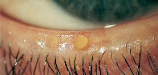
Disturbed Sensory Perception (visual) related to Blepharitis


Disturbed Sensory Perception (visual) related to Blepharitis
Nursing Care Plan for Blepharitis

Blepharitis or better known as inflammation of the eyelids is one of the eye diseases to watch out for. The disease is caused by the oil glands at the base of the lashes were damaged, causing itching in the eyelids, irritation, even to inflammation. If this is allowed then it would be blepharitis.

Although blepharitis does not cause permanent damage to the eye, but very disturbing activities of our vision.

Symptoms of Blefarits as follows:
  • The eyelids are often greasy.
  • Itch on the eyelids.
  • Burned in the eye.
  • Eyes look red.
  • Frequent watery eyes.
  • Eye swelling of the eyelids.
  • Eyelash gross waking.
  • Very sensitive to light.
  • Have peeling skin around the eyes.
  • Eyelashes fall out.
  • Eyelashes abnormal and irregular direction.

Main causes of the occurrence of blepharitis (inflammation of the eyelids)
  • Infected by the bacteria.
  • Glands damaged eye.
  • Dandruff of the scalp to eyebrows.
  • Rosacea.
  • Allergic to something eg eye drops, eye lens.


Nursing Diagnosis and Interventions for Blepharitis :

Disturbed Sensory Perception (visual) related to reception interference status sensory organs.

Goal: Increase the visual acuity within the limits of individual situations.

Expected outcomes:
  • Know the sensory disturbances and compensate for changes.
  • Identify / fix potential hazards in the environment.

Intervention:

1) Determine the visual acuity, note whether one or both eyes are involved.
Rational: individual needs and choice of interventions varied causes vision loss occurs slowly and progressively.

2) Observe the signs and symptoms of disorientation.
Rational: woke up in unfamiliar surroundings and have limited vision.

3) Orient the patient on the environment, other people in the area.
Rationale: provides increased comfort and familiarity.

4) Notice about blurred vision and eye irritation, which can occur when using eye drops.
Rational: impaired vision / irritation can end 1-2 hours after use of eye drops, gradually decreases with usage.

5) Place the items needed / call bell within reach positions on the near side.
Rational: allowing patients to see objects more easily and facilitate the call for help when needed.

Ineffective Airway Clearance - NCP for Bronchiectasis

Nursing Care Plan for Bronchiectasis

Bronchiectasis is a chronic dilatation of the bronchi and bronchioles that may be caused by various conditions, including lung infections and bronchial obstruction; foreign body aspiration, vomit, and objects from the upper respiratory tract; and the pressure due to a tumor, blood vessels dilated and enlarged lymph nodes (Brunner & Suddart, 2002).

According Suyono (2001) etiology of bronchiectasis are:

1. Infection
Bronchiectasis often occur after a child suffering from pneumonia who frequently relapse and long lasting. Pneumonia is generally a pertussis or influenza complications suffered during the child, pulmonary tuberculosis, and so on.

2. Abnormalities hereditary or congenital abnormalities
In this case bronchiectasis occurs in the womb. Genetic factors or growth factors and fetal development plays an important role. Usually has the characteristics on almost all branches of the bronchi in the lungs one or two. Usually accompanied by other congenital diseases.

3. Bronchial obstruction
Obstruction is meant as a corpus alienum, bronchial carcinoma and other external pressure against the bronchi.


Pathophysiology

According to Brunner & Suddarth (2002) pathophysiology of bronchiectasis starting from infections that damage the bronchial wall, causing loss of supporting structure and produce thick sputum that can eventually clog the bronchi. Bronchial walls become stretched permanently as a result of severe coughing, infection extends to peri bronchial, so that in the case of secular bronchiectasis, each tube is actually dilated pulmonary abscess, which exudates to flow freely through the bronchi. Bronchiectasis is usually local, attacking lung lobe segment. Lower lobe most often affected.

Retention secretion and obstruction resulting eventually cause obstruction distal side alveoli collapse (atelectasis). Due to scarring or fibrosis replaces lung tissue inflammatory reaction that function. At the time the patient suffered respiratory insufficiency with a decrease in vital capacity, decreased ventilation, and an increase in the ratio of residual volume to total lung capacity. There is damage to the gas mixture in the inspiration (ventilation-perfusion mismatch) and hypoxemia.


According Suyono (2001) the signs and symptoms of bronchiectasis as follows:

1. Cough
Hemoptysis is characterized, among others; ongoing chronic productive cough, sputum amount varies, generally polynomial in the morning after there is a sleeping position or wake up from sleep. Sputum consists of three layers:
  • The top layer; rather cloudy, consisting of mucus.
  • The middle layer; clearly consists of saliva.
  • The bottom layer; turbid, consisting of pus and tissue necrosis of bronchial damaged.
2. Hemoptysis
Caused by necrosis or destruction of the bronchial mucosa blood vessels (rupture) and the resulting bleeding.

3. Shortness of breath (dyspnea)
Onset of shortness of breath depends on the extent of bronchiectasis, sometimes causing wheezing sound due to bronchial obstruction.

4. Recurrent fever
Bronchiectasis is a chronic disease, often experience recurrent infection of the bronchi and the lungs, often resulting in fever (recurrent fever).

5. Physical Abnormalities
  • Cyanosis
  • Clubbing
  • Bronchi wet
  • Whezing

Nursing Care Plan for Bronchiectasis

Nursing Diagnosis : Ineffective airway clearance related to the increased production of secretions, thick secretions.

Goal: Maintain a patent airway with breath sounds clean / clear.

Expected outcomes: Demonstrate behaviors to improve airway clearance (effective cough, and issued a secret.)

Intervention:
1. Auscultation of breath sounds and record their breath sounds.
R /: The degree of bronchospasm occurs with airway obstruction and can / not characterized by the presence of breath sounds.

2. Assess / monitor respiratory frequency. Note the ratio of inspiration and expiration.
R /: Tacipneu common to some degree can be found at the reception or during stress / acute infection process. Slowed breathing and elongated compared inspiration expiration frequency.

3. Assess the patient to a comfortable position, Height head of the bed and sat on the back of the bed.
R /: Elevation headboard ease respiratory function by means of gravity. And make it easier to breathe, and help decrease muscle weakness and can be as a tool chest expansion.

4. Help abdominal breathing exercises or lips.
R / To cope with and control of dyspnea and lower air entrapment.

5. Observe the characteristic cough and aid effectiveness action to cough effort.
R /: Knowing the effectiveness of cough.

6. Increase fluid intake till 3000ml / day as tolerated heart and give a warm and fluid intake between as a meal replacement.
R /: Hydration helps to lower the viscosity of secretions, simplify expenditure warm fluids can reduce bronchospasm. Liquids between meals can increase gastric distension and pressures diaphragm.

7. Give the drug as indicated.
R /: Speed up the healing process.

Nursing Diagnosis and Interventions for Dehydration


Nursing Diagnosis for Dehydration
  1. Fluid volume deficit related to excessive output, less intake.
  2. Risk for ineffective tissue perfusion related to decreased blood flow.
  3. Risk for impaired skin integrity related to decreased skin turgor.
  4. Activity intolerance related to physical weakness.
  5. Risk for Decreased cardiac output related to a decrease in systemic vascular resistance.


Nursing Care Plan for Dehydration

Nursing Interventions for Dehydration

1. Fluid volume deficit related to excessive output, less intake.

Goal: adequate fluid volume so that fluid volume deficiency can be overcome.

Expected outcomes:
  • Maintain fluid balance.
  • Vital signs (pulse = 80-100 beats / min, temperature = 36-37oC)
  • Capillary refill less than 3 seconds.
  • Akral warm.
  • Urine output: 1-2 cc / kg body weight / hour.

Intervention:
  • Monitor vital signs, capillary refill, the status of the mucous membranes.
  • Discuss strategies to stop vomiting and use of laxatives / diuretics.
  • Identification of a plan to increase the optimal fluid balance.
  • Assess the results of the test function electrolyte / kidney.
  • Give / supervise administration of IV fluids.
  • Additional potassium, oral or N as indicated.

2. Risk for ineffective tissue perfusion related to decreased blood flow.

Goal: Maintain / improve tissue perfusion.

Expected outcomes:
  • Vital signs are stable BP = 120/80 mmHg, pulse = 80-100 beats / min, no pale skin.
  • Warm skin.
  • Palpable peripheral pulses.
  • Adequate urine output from 0.5 to 1.5 cc / kg / body weight.
  • CRT is less than 2 seconds.
  • Composmentis consciousness.
  • No chest pain.

Intervention:
  • Assess changes in the level of consciousness, dizziness complaints.
  • Auscultation apical pulse, watch heart rate / rhythm.
  • Assess the skin against the cold, pale, sweating.
  • Record output and urine specific gravity.
  • Observation pale skin, redness, change positions frequently.
  • Keep an eye on pulse oximetry.
  • Give IV fluids as indicated.


3. Risk for impaired skin integrity related to decreased skin turgor.

Goal: Identify and maintain the skin smooth, supple, intact.

Expected outcomes:
  • Good skin turgor, skin intact, no blisters, no redness.

Intervention:
  • Observation reddish, pale.
  • Use skin cream.
  • Discuss the importance of changes in position, it is necessary to maintain the activity.
  • Emphasize the importance of nutrient input / adequate fluid.

Nursing Diagnosis, Definition, Outcomes and Interventions - Risk for Infection


Risk for Infection related to the invasion of microorganisms in the body

Goal : after the act of nursing for 3x24 hours of infection did not occur.

Expected outcomes:
  • Patients will show a careful hand-washing techniques.
  • Patients will be free of the nosocomial infection during hospitalization.
  • Patients will demonstrate knowledge of the risk factors associated with infection and appropriate precautions to prevent infection.

Intervention - Risk for Infection:

1. Monitor for signs and symptoms of infection.
R /: To determine whether there is an infectious process.

2. Monitor laboratory results, Monitor the patient's temperature.
R /: Leukocyte increased and increased body temperature is not expected, a sign of infection.

3. Use antiseptic technique when taking action to clients.
R /: Prevent cross-infection.

4. Emphasize the need to wash hands regularly / thoroughly before and when handling food, after toileting.
R /: Many viruses such as cytomegalovirus (CMV) can be excreted in the urine for more than 4 years after exposure and possibly transmitted through poor hygienic.

5. Encourage clients to drink 6 to 8 glasses of fluid every day. Discuss the role of acidic residues in the diet and add juice or orange cranberr.
R /: Helps to acidify the urine and help prevent UTIs.

6. Encourage clients to try Kegel exercises (tightening the perineum) throughout the day.
R /: Fix support for pelvic organs, strengthen and increase the elasticity of the pubococcygeus muscle, better control urination.

7. Encourage the use of cotton underwear, and avoid using bath tub, if a client has a history of Urinary Tract Infections (UTIs).
R /: Static urinary and glycosuria may mempredisposisikan prenatal clients on a urinary tract infection or UTI, especially when history include urinary problems / kidney.

8. Get a routine urine sample for microscopic examination, pH, presence of leukocyte cells, and also culture and sensitivity, according to the indication. Report the number of colonies greater than one hundred thousand per milliliter.
R /: This is to detect the presence of microorganisms in the body. This is to detect the presence of microorganisms in the body. High leukocyte cell count is an indicator of infection.

9. Instruct the patient to always clean the areas that are reddish.
R /: Prevent the entry of other bacteria that can cause infection.

10. Collaboration with the medical team to provide antibiotics.
R /: Antibiotics can help fight infection.